Cells of the Nervous System
Tareq Yousef
The Neuron Doctrine: A Turning Point in Brain Science
Today, psychologists and neuroscientists can study the brain with advanced tools such as brain imaging and electrophysiology, allowing them to observe neurons in action. But this modern knowledge is built on earlier discoveries that first revealed what neurons are.
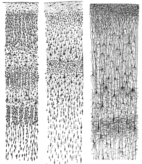
In the late 19th century, scientists were still debating what the brain was made of. Many believed it was a continuous network of tissue, like a giant web. This view began to change when Camillo Golgi developed a special silver staining technique that revealed the detailed structure of individual cells in the nervous system. Building on this method, Santiago Ramón y Cajal demonstrated that the brain is not a continuous mass, but is instead made up of distinct cells, what we now call neurons. Cajal’s work laid the foundation for the neuron doctrine, the idea that neurons are the basic units of the nervous system. This discovery transformed neuroscience and provided the starting point for understanding how thought, behavior, and experience arise from neural activity.
Modern Understanding of the Human Brain
Psychologists striving to understand the human mind may study the nervous system. Learning how the body’s cells and organs function can help us understand the biological basis of human psychology. The nervous system is composed of two basic cell types: glia and neurons. Glia (a single one is a glial cell) are traditionally thought to play a supportive role to neurons, both physically and metabolically (i.e., supplying nutrients and maintaining their function). Glia provide scaffolding on which the nervous system is built, help neurons line up closely with each other to allow neuronal communication, provide insulation to neurons, transport nutrients and waste products, and mediate immune responses.
For years, researchers believed that there were many more glia than neurons; however, more recent work from Suzanna Herculano-Houzel’s laboratory has called this long-standing assumption into question and has provided important evidence that there may be a nearly 1:1 ratio of glia to neurons. This is important because it suggests that human brains are more similar to other primate brains than previously thought (Azevedo et al, 2009; Hercaulano-Houzel, 2012; Herculano-Houzel, 2009). Neurons, on the other hand, serve as interconnected information processors that are essential for all of the tasks of the nervous system. This means that these cells can act as different parts of a machine (the entire nervous system) to carry out a function (like moving your leg, or retrieving a memory). This section briefly describes the structure and function of neurons.
Neuron Structure
Neurons are the central building blocks of the nervous system, 100 billion strong at birth. Like all cells, neurons consist of several different parts, each serving a specialised function (Figure BB.2). A neuron’s outer surface is made up of a semipermeable membrane. The semipermeable nature of the membrane allows smaller molecules and molecules without an electrical charge to pass through it, while stopping larger or highly charged molecules.
, axon, and terminal buttons. A myelin sheath covers part of the neuron.")
The nucleus of the neuron is located in the soma, or cell body. The soma has branching extensions known as dendrites. The neuron is a small information processor, and dendrites serve as input sites where signals are received from other neurons. These signals are transmitted electrically across the soma and down a major extension from the soma known as the axon, which ends at multiple terminal buttons (also commonly referred to as boutons–the French word for buttons). The terminal buttons contain synaptic vesicles that house neurotransmitters, the chemical messengers of the nervous system.
Axons range in length from a fraction of an inch to several feet. In some axons, glia form a fatty substance known as the myelin sheath, which coats the axon and acts as an insulator, increasing the speed at which the signal travels. The myelin sheath is not continuous and there are small gaps that occur down the length of the axon. These gaps in the myelin sheath are known as the Nodes of Ranvier. The myelin sheath is crucial for the normal operation of the neurons within the nervous system; the loss of the insulation it provides can be detrimental to normal function.
To understand how this works, let’s consider an example. Phenylketonuria (fen-ul-key-toe-NU-ree-uh), also called PKU, causes a reduction in myelin and abnormalities in white matter cortical and subcortical structures (regions of the brain below the surface). This disorder is associated with a variety of issues including severe cognitive deficits (i.e., difficulties with everyday functioning or learning), exaggerated reflexes, and seizures (Anderson & Leuzzi, 2010; Huttenlocher, 2000). Another disorder, multiple sclerosis (MS), an autoimmune disorder, involves a large-scale loss of the myelin sheath on axons throughout the nervous system. The resulting interference in the electrical signal prevents the quick transmission of information by neurons and can lead to a number of symptoms, such as dizziness, fatigue, loss of motor control, and sexual dysfunction. While some treatments may help to modify the course of the disease and manage certain symptoms, there is currently no known cure for multiple sclerosis.
Watch this video: Tricky Topics: Neuronal Structure (17.5 minutes)
“Tricky Topics: Neuronal Structure” video by FirstYearPsych Dalhousie is licensed under the Standard YouTube Licence.
Here is the Tricky Topics: Neuronal Structure transcript.
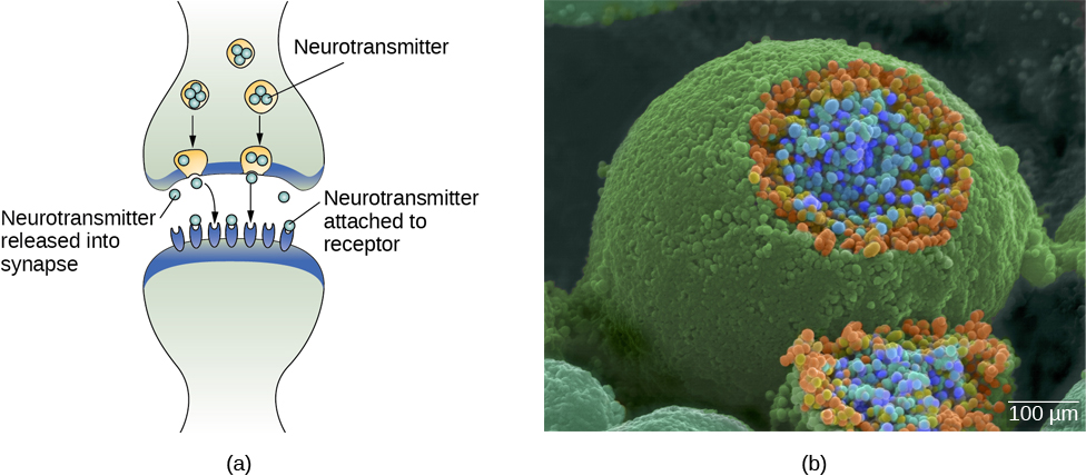
In typically functioning nervous systems, the neuronal signal moves rapidly down the axon to the terminal buttons, where synaptic vesicles release neurotransmitters into the synaptic cleft (Figure BB.3). The synaptic cleft is a very small space between two neurons and is an important site where communication between neurons occurs. Once neurotransmitters are released into the synaptic cleft, they travel across it and bind with corresponding receptors on the dendrite of an adjacent neuron. Receptors, proteins on the cell surface where neurotransmitters attach, vary in shape, with different shapes matching different neurotransmitters.
How does a neurotransmitter know which receptor to bind to? The neurotransmitter and the receptor have what is referred to as a lock-and-key relationship — specific neurotransmitters fit specific receptors similar to how a key fits a lock. The neurotransmitter binds to any receptor that it fits.
Watch this video: Tricky Topics: Synaptic Transmission (9 minutes)
“Tricky Topics: Synaptic Transmission” video by FirstYearPsych Dalhousie is licensed under the Standard YouTube Licence.
Here is the Tricky Topics: Synaptic Transmission transcript.
Neuronal Communication
Now that we have learned about the basic structures of the neuron and the role that these structures play in neuronal communication, let’s take a closer look at the signal itself — how it moves through the neuron and then jumps to the next neuron, where the process is repeated.
We start our journey at the neuronal membrane. The neuron exists in a fluid environment; it is surrounded by fluid outside the cell, called extracellular fluid, and contains fluid inside the cell, known as intracellular fluid or cytoplasm. The neuronal membrane acts as a barrier, keeping these two fluids separate. This role is crucial because the electrical signal passing through the neuron relies on the electrical difference between the intra- and extracellular fluids. This charge difference, called the membrane potential, supplies the energy for the signal.
The electrical charge of the fluids is caused by charged molecules (ions) dissolved in the fluid. The semipermeable nature of the neuronal membrane somewhat restricts the movement of these charged molecules, and, as a result, some of the charged particles tend to become more concentrated either inside or outside the cell.
Between signals, the neuron membrane’s potential is held in a state of readiness , called the resting potential. The typical resting potential of a neuron is -70 millivolts (mV). Like a rubber band stretched out and waiting to spring into action, ions line up on either side of the cell membrane, ready to rush across the membrane when the neuron goes active and the membrane opens its gates (i.e., a sodium-potassium pump that allows movement of ions across the membrane). Ions in high-concentration areas are ready to move to low-concentration areas in a process called osmosis (you may have come across the term before), and positive ions are ready to move to areas with a negative charge. This is a fundamental process, where negatively charged particles are attracted to positive charges, just like the interaction of two magnets.
In the resting state, sodium (Na+) is at higher concentrations outside the cell, so it will tend to move into the cell. Potassium (K+), on the other hand, is more concentrated inside the cell, and will tend to move out of the cell (Figure BB.4). In addition, the inside of the cell is slightly negatively charged compared to the outside. This difference in charge, known as the electrochemical gradient, acts on sodium, causing it to move into the cell.

From this resting potential state, the neuron receives a signal and its state changes abruptly (Figure BB.5). When a neuron receives signals at the dendrites — due to neurotransmitters from an adjacent neuron binding to its receptors — small pores, or gates, open on the neuronal membrane, allowing Na+ ions, propelled by both charge and concentration differences, to move into the cell. With this influx of positive ions, the internal charge of the cell becomes more positive. If that charge reaches a certain level, called the threshold of excitation, usually around the neuron becomes active and the action potential begins.
Several additional pores open, leading to a massive influx of positively charged sodium ions creating a significant spike in the membrane potential, the peak of the action potential. At this peak, the sodium gates close, and the potassium gates open. Positively charged potassium ions start to exit the cell rapidly, initiating the process of repolarization. Initially, the cell becomes more negative than its resting potential, known as hyperpolarization, and then it stabilises, returning to its resting potential.

This positive spike represents the action potential: the electrical signal that typically moves from the cell body down the axon to the axon terminals. The electrical signal moves down the axon with the impulses jumping in a leapfrog fashion between the Nodes of Ranvier. The Nodes of Ranvier are natural gaps in the myelin sheath. At each point, some of the sodium ions that enter the cell diffuse to the next section of the axon, raising the charge past the threshold of excitation and triggering a new influx of sodium ions. The action potential moves all the way down the axon in this way until reaching the terminal buttons.
The action potential is an all-or-none phenomenon. In simple terms, this means that an incoming signal from another neuron is either sufficient or insufficient to reach the threshold of excitation. There is no in-between, and there is no turning off an action potential once it starts. Think of it like sending an email or a text message. You can think about sending it all you want, but the message is not sent until you hit the send button. Furthermore, once you send the message, there is no stopping it.
Since it’s an all-or-none response, the action potential is reproduced, or propagated, at its full strength along every point of the axon. Imagine it like a lit fuse on a firecracker — it doesn’t lose intensity as it travels down the axon. This all-or-none property explains why your brain perceives an injury to a distant body part, such as your toe, as equally painful as one to your nose (which is closer to your brain).
As noted earlier, when the action potential arrives at the terminal button, the synaptic vesicles release their neurotransmitters into the synaptic cleft. The neurotransmitters travel across the synapse and bind to receptors on the dendrites of the adjacent neuron, and the process repeats itself in the new neuron (assuming the signal is sufficiently strong to trigger an action potential). Once the signal is delivered, excess neurotransmitters in the synaptic cleft drift away, break down into inactive fragments (degradation), or are reabsorbed in a process known as reuptake.
Reuptake involves the neurotransmitter being pumped back into the neuron that released it, in order to clear the synapse (Figure BB.6). Clearing the synapse serves both to provide a clear “on” and “off” state between signals and to regulate the production of neurotransmitter (full synaptic vesicles signal that no additional neurotransmitters need to be produced). The synapse can also be cleared via degradation of the neurotransmitter, which typically involves an enzyme breaking the neurotransmitter down into its components, so that it can no longer interact with the receptors on the postsynaptic neuron.

Neuronal communication is often referred to as an electrochemical event. The movement of the action potential down the length of the axon is an electrical event, and movement of the neurotransmitter across the synaptic space represents the chemical portion of the process. However, there are some specialised connections between neurons that are entirely electrical. In such cases, the neurons are said to communicate via an electrical synapse. In these cases, two neurons physically connect to one another via gap junctions that allow the current from one cell to pass into the next. There are far fewer electrical synapses in the brain, but those that do exist are much faster than the chemical synapses that have been described above (Connors & Long, 2004).
Watch this video: Tricky Topics: Action Potentials (18 minutes)
“Tricky Topics: Action Potentials” video by FirstYearPsych Dalhousie is licensed under the Standard YouTube Licence.
Here is the Tricky Topics: Action Potentials transcript.
Neurotransmitters
Neurons communicate with one another by releasing chemical messengers called neurotransmitters. Different neurotransmitters are linked to different functions in the brain and body (see Table BB.1). For example, acetylcholine plays an important role in muscle movement, learning, memory, and arousal(Picciotto et al., 2012). Dopamine is associated with reward, motivation, and motor control. Serotonin influences mood, sleep, and appetite (Ko & Strafella, 2012), while gamma-aminobutyric acid (GABA) acts as the brain’s main inhibitory neurotransmitter, helping to calm neural activity (Bowery & Smart, 2006). In contrast, glutamate is the primary excitatory neurotransmitter and is essential for learning and memory (Zhou & Danbolt, 2014). By working together in complex systems, these neurotransmitters regulate a wide range of psychological and physiological processes, from movement and arousal to emotion and cognition.
| Neurotransmitter | Involved in | Potential effect on behaviour |
|---|---|---|
| Acetylcholine | Muscle activity, memory | Increased arousal, enhanced cognition |
| Beta-endorphin | Pain, pleasure | Decreased anxiety, decreased tension |
| Dopamine | Mood, sleep, learning | Increased pleasure, suppressed appetite |
| Gamma-aminobutyric acid (GABA) | Inhibitory control, sleep | Decreased anxiety, decreased tension |
| Glutamate | Memory, learning | Increased learning, enhanced memory |
| Norepinephrine | Heart function, intestinal function, alertness | Increased arousal, suppressed appetite |
| Serotonin | Mood, sleep | Modulated mood, suppressed appetite |
Glial Cells:
(i.e., glia) Cells that surround and link to the neurons, protecting them, providing them with nutrients, and absorbing unused neurotransmitters.
Neuron:
A cell in the nervous system that is responsible for receiving and transmitting information.
Soma:
A cell body that contains the nucleus of the cell and keeps the cell alive.
Axon:
A long, segmented fibre within the neuron that transmits information away from the cell body toward other neurons or to the muscles and glands.
Myelin Sheath:
A layer of fatty tissue surrounding the axon of a neuron that both acts as an insulator and allows faster transmission of the electrical signal.
Node of Ranvier:
A series of breaks between the sausage-like segments of the myelin sheath that allow electrical charges to move down the axon.
Resting Potential:
A state in which the interior of the neuron contains a greater number of negatively charged ions than does the area outside the cell.
Action Potential:
The change in electrical charge that occurs in a neuron when a nerve impulse is transmitted.
Reuptake:
A process in which neurotransmitters that are in the synapse are reabsorbed into the transmitting terminal buttons, ready to again be released after the neuron fires.
